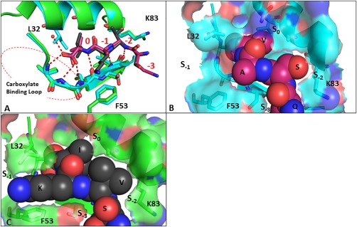
Figure 2.

(A) Comparison of binding modes of Class I PKCα (QSAV (magenta)) peptide with Class II GluA2 (SVKI (black (PBD:3HPK))) with PICK1. Space filling representation of the PKCα (B) and GluA2 (C) peptides on the surface of PICK1. An interactive view is available in the electronic version of the article
