Figure 3.

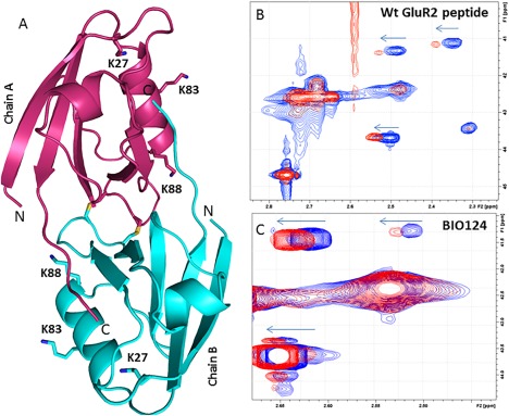
(A) Overall Structure of PICK1 PDZ‐QSAV with Lysines 6Å away from peptide binding pocket labeled. Shifts of methylated Lysines in 2D Protein NMR for (B) Wt GluR2 peptide and (C) BIO124.

(A) Overall Structure of PICK1 PDZ‐QSAV with Lysines 6Å away from peptide binding pocket labeled. Shifts of methylated Lysines in 2D Protein NMR for (B) Wt GluR2 peptide and (C) BIO124.