
Figure 3.
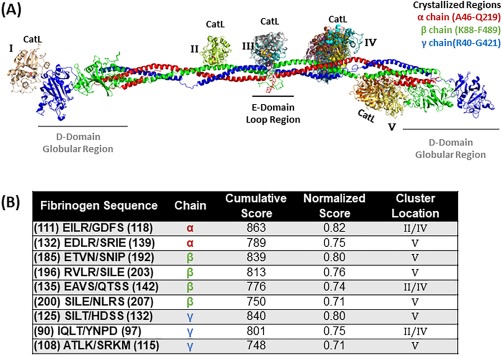
Top scored locations for cathepsin L binding and cleavage of fibrinogen. (A) Molecular docking of cathepsin L on fibrinogen monomer with the α chain is in red, the β chain is in green, and the γ chain is in blue. Cathepsin L binds in various formations and each color indicates a different docking model (note there is some overlap with the model found in cluster location B and D). Structures used were PDB ID 3GHG (fibrinogen) and 5F02 (cathepsin L). (B) Predicted fibrinogen cleavage sites by cathepsin L from PACMANS and molecular docking models.