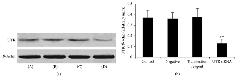
Figure 1.
The expression of UTR protein in rat myocardium after UTR siRNA in vivo transfection. (a) UTR protein was detected by Western blot. β-Actin was used as the loading control. (b) Densitometric quantification of UTR protein expression. (A): control; (B): negative; (C): transfection reagent; and (D): UTR siRNA. Results are expressed as mean ± SD (n = 5, each group). ∗∗p < 0.01 versus control group.

