
Figure 3. FGF1 prevents DN in STZ-induced T1D mice.
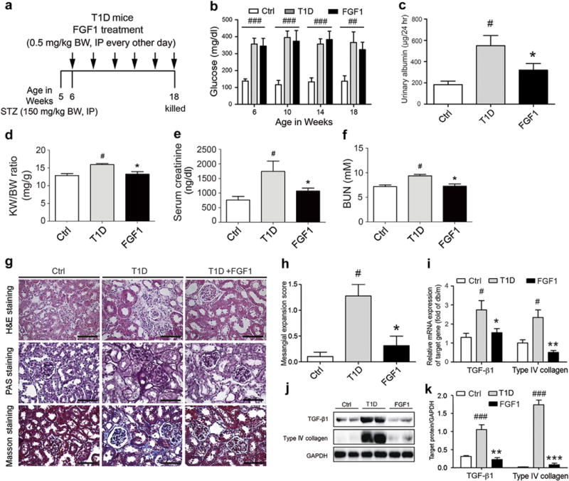
(a) FGF1 treatment protocol for mice with T1D. Arrows indicate that mice received i.p. injections of vehicle or FGF1 (0.5 mg/kg BW) for 12 weeks and then were killed at 18 weeks for study. (b,c) Blood glucose level (nonfasted) measured weekly and 24-hour urinary albumin excretion from the normal control (Ctrl), T1D, and T1D+FGF1 groups. The kidney-BW ratio (d), serum creatinine (e), and BUN (f) in mice from the Ctrl, T1D, and FGF1 (T1D+FGF1) groups. Mice received i.p. injections of 0.5 mg/kg of FGF1 or physiologic saline every other day for 12 weeks and were evaluated for biometric and biochemical parameters. Data are shown as the mean ± SEM; #P < 0.05 versus vehicle control; *P < 0.05 versus T1D mice (N = 7–9). (g) Representative images of renal tissue stained with H&E, PAS (indicating glycogen) for evaluation of mesangial expansion, and Masson trichrome for type IV collagen. Bar 100 = μm. (h) The glomerular mesangial expansion score determined from histology sections. (i) Gene expression of transforming growth factor β1 and type IV collagen in renal tissue from the Ctrl, T1D, and T1D+FGF1 groups. (j) Western blot analysis of TGF-β1 and type IV collagen in renal tissue from the Ctrl, T1D, and T1D+FGF1 groups. GAPDH expression was used for normalization of protein loading. (k) Quantification of Western blot by densitometric analysis. (b-k) Data are shown as the mean ± SEM; #P < 0.05, ##P < 0.01, and ###P < 0.001 versus Ctrl; *P < 0.05, **P < 0.01, and ***P < 0.001 versus T1D mice (N = 7–9). BUN, blood urea nitrogen; BW, body weight; Ctrl, control; FGF1, fibroblast growth factor 1; GAPDH, glyceraldehyde-3-phosphate dehydrogenase; H&E, hematoxylin and eosin; KW, kidney weight; PAS, periodic acid–Schiff; STZ, streptozotocin; T1D, type 1 diabetes; TGF-1β, transforming growth factor 1β. To optimize viewing of this image, please see the online version of this article at www.kidney-international.org.