
Figure 5. FGF1 prevents HG–induced inflammatory responses in renal mesangial cells.
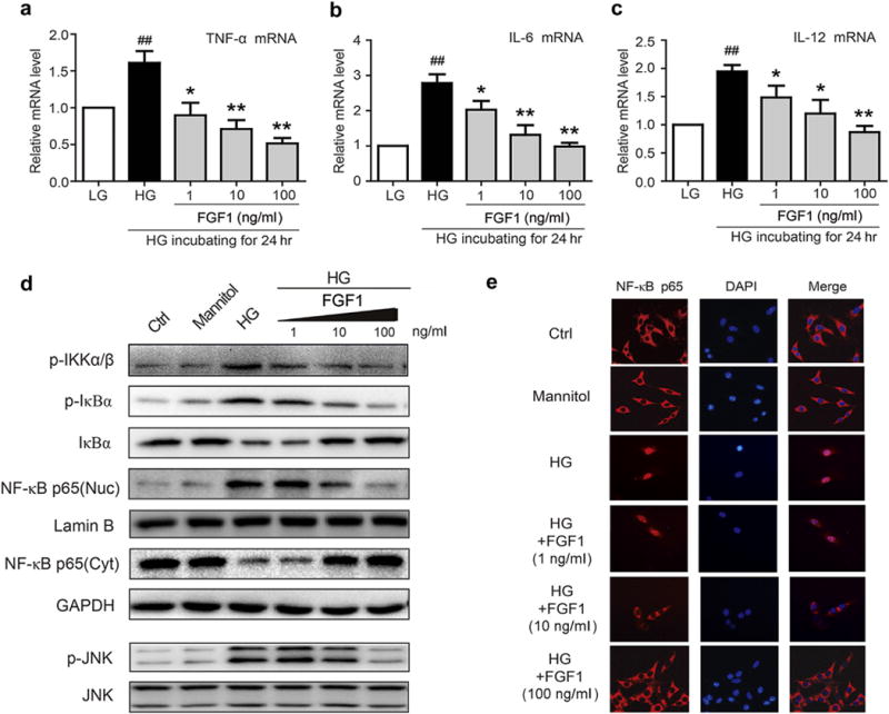
(a–c) Mesangial cells were pretreated with FGF1 (1, 10, or 100 ng/ml) for 2 hours, followed by treatment with HG (25 mM) for an additional 24 hours, and mRNA of proinflammatory cytokines, TNF-β, IL-6, and IL-12 determined by reverse transcriptase polymerase chain reaction; values normalized to the housekeeping gene GAPDH and reported as the mean ± SEM. ##P < 0.01 versus LG control; *P < 0.05, **P <.01 versus HG; N = 3. (d) Effects of FGF1 pretreatment (1, 10, or 100 ng/ml for 1 hour) of mesangial cells on HG-induced (2 hours) activation of NF-κB and JNK. (e) Immunofluorescent localization of NF-κB p65 subunit (red) in mesangial cells: Ctrl, mannitol (osmotic control), HG, HG+FGF1 with the indicated concentration; nuclei stained with fluorescent DAPI (blue). Ctrl, control; DAPI, 4′,6-diamidino-2-phenylindole; FGF1, fibroblast growth factor 1; GAPDH, glyceraldehyde-3-phosphate dehydrogenase; HG, high glucose; JNK, c-Jun N-terminal kinase; LG, low glucose; NF-κB, p-JNK, phosphorylation of c-Jun N-terminal kinase; RT-PCR, reverse transcriptase polymerase chain reaction; TNF-α, tumor necrosis factor-α. To optimize viewing of this image, please see the online version of this article at www.kidney-international.org.