
Fig. 1.
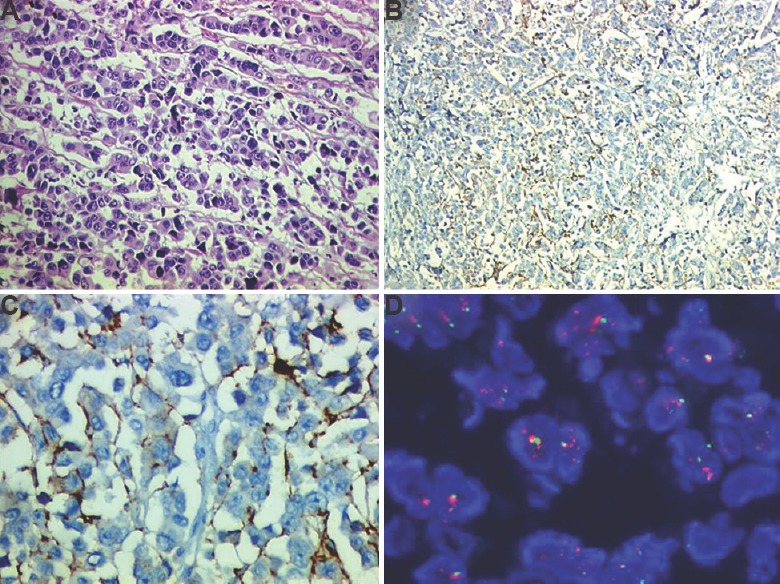
A patient affected by gap in biomarker testing (A) haematoxylin and eosin section confirmed that tumour was poorly fixed (H & E: 200×) (B) weak HER2neu staining due to poor fixation (immunoperoxidase: 200×), (C) higher power to indicate that HER2neu would be interpreted as score 1+/negative (immunoperoxidase: 400×), and (D) tumour as tested by fluorescent in situ hybridization found to be HER2neu amplified (Oil).