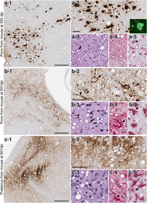
Fig. 2.

Immunohistochemistry and neuropathology of tg66 mice injected with Y226X human brain homogenate. Panel a Pons region of a mouse euthanized at 593 dpi. PrPSc was detected by IHC using biotinylated antibody 3F4 as described in the methods (panel a-1). Large and medium-sized PrPSc deposits are seen at higher magnification (a-2). Inset in a-2 shows Thioflavin S staining of one aggregate. Typical prion disease vacuolation (arrow) is shown by H&E staining (a-3), and astrogliosis and microgliosis (arrow) are shown by anti-GFAP staining (a-4) and anti-Iba1 staining (a-5). Panel b Pons region of a mouse euthanized at 601 dpi. PrPSc staining showed smaller coarse deposits (b-1), and perineuronal and linear axonal staining (arrows) could be seen at higher magnification (b-2). Vacuolation, astrogliosis and microgliosis (arrows) was also prominent in this same area (b-3, b-4, b-5). Panel c: Thalamus of same mouse shown in panel b showed slightly finer staining of PrPSc at both low (c-1) and high (c-2) magnification. Prominent vacuolation (c-3), astrogliosis (c-4) and microgliosis (c-5) was also noted (arrows). Scale bars shown in a-1, b-1 and c-1 are 200 μm, scale bars shown in a-2, b-2 and c-2 are 50 μm and apply to each subsequent panel within the same figure letter
