
Fig. 3.
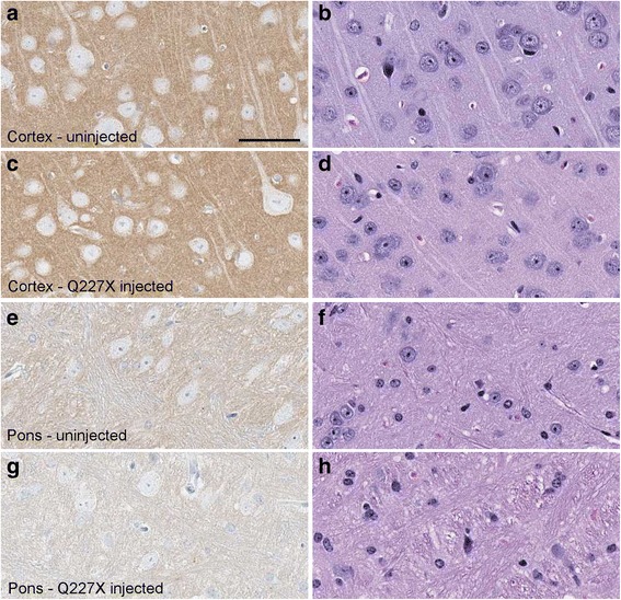
Histology and immunohistochemical staining of PrP in two brain regions of a tg66 mouse injected with Q227X brain homogenate at 743 dpi, and an uninfected aged control tg66 mouse (age 649 days). Panels a, c, e, g show PrP staining with biotinylated-3F4 antibody, and panels b, d, f, h show H&E staining. Uninfected cortex (a, b), Q227X injected cortex (c, d), uninfected pons (e, f), Q227X-injected pons (g, h). Panels a and c show darker tan staining than panels e and g due to a higher level of background PrPC in cortex compared to pons. No prion disease vacuoles or significant deposits suggestive of PrPSc were observed in uninfected or the Q227X-injected mice. Scale bar in panel a is 50 μm and is valid for all panels