Fig. 5.

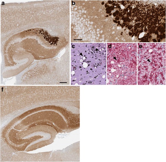
Immunohistochemistry and neuropathology of a G131V-injected tg66 mouse at 731 days post-injection. a-e Oriens layer of dorsal hippocampus. Coarse plaque-like staining of PrPSc by antibody 3F4 is seen at low (a) and high (b) magnification. Rectangular box in a denotes area seen in b. In the same area, gray matter vacuoles (arrow) were noted by H&E staining (c), and astrogliosis (d) and microgliosis (e) were observed (arrows) by IHC using antibodies to GFAP and Iba1. Panel f: No PrPSc deposits were seen in a control age-matched uninfected mouse in the same hippocampal region shown in (a) The scale bar in panel a is 200 μm and applies to panel f. In b the bar is 50 μm and applies to panels b-e
