Figure 3.

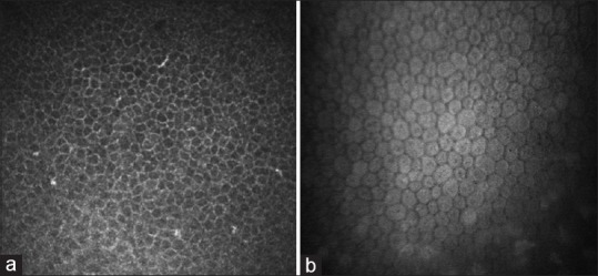
Image of epithelium (a) and endothelium (b) in healthy eye. Epithelium image was captured with Heidelberg Retina Tomograph II (Heidelberg, Germany) and endothelium image with Confoscan4 (Nidek Technologies, Japan)

Image of epithelium (a) and endothelium (b) in healthy eye. Epithelium image was captured with Heidelberg Retina Tomograph II (Heidelberg, Germany) and endothelium image with Confoscan4 (Nidek Technologies, Japan)