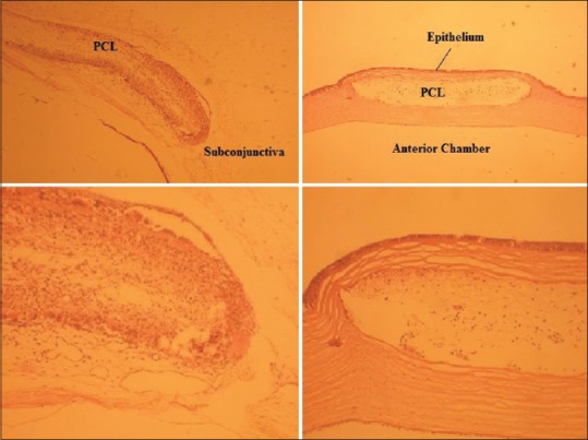
Figure 3.

Routine histopathologic slides of the subconjunctival patch (upper left) and corneal stromal patch (upper right) in rabbit A and their magnified views (lower left and lower right, respectively). Images illustrate the infiltration of host cells through nanofibers
