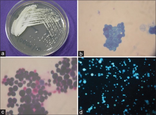
Figure 2.

Macroscopic and microscopic appearance of Prototheca wickerhamii isolated from corneal scraping. (a) Colony morphology of P. wickerhamii on sabouraud dextrose agar. (b-d) Methylene blue stain (×100), Gram stain (×100), and potassium hydroxide/calcofluor (×20) stained preparations showing the oospores of Prototheca wickerhamii
