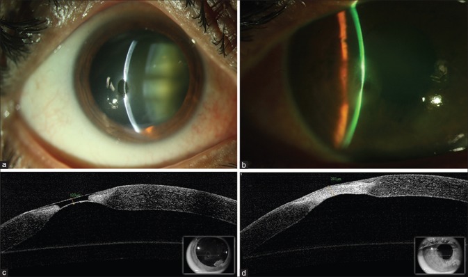
Figure 1.
(a) Slit lamp photograph of the right eye showing significant thinning paracentrally. (b) High-resolution optical coherence tomography at the site of corneal melt and descemetocele. (c) Slit lamp photograph at 6 weeks showing scarring and increased stromal thickness at the site of melt. (d) High-resolution optical coherence tomography showing the healed area with epithelization and incorporation of amniotic membrane at the site of descemetocele. The stromal thickness measured 281 μ. The line scan (inset) shows the same area of imaging as obtained at baseline before amniotic membrane transplantation

