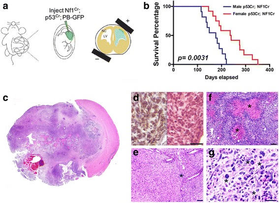
Fig. 1.

Deletion of neurofibromin and p53 in vivo results in sexually dimorphic gliomagenesis. a Schematic of bioelectroporation strategy. The uterus is delivered intact to the external environment (left hand panel). The cerebral ventricles are identified in each pup by trans-illumination and injected with gRNAs directed against neurofibromin and p53 (middle panel). An electric field is imposed across the head of each pup to drive the gRNAs into the sub-ventricular region (right hand panel). b Survival was significantly shorter for male mice compared to female mice. While all mice succumbed to tumors, median survival for male mice (n = 11) was 176 days and for female mice (n = 14), 238 days. p = 0.0031 as determined by log-rank test of Kaplan-Meier survival curves. c Large invasive tumors formed in all mice. d Tumors were diagnosed as astrocytomas based on their expression of glial fibrillary acidic protein (GFAP, brown-right). Corresponding H&E (right) and GFAP (left) staining from a CRISPR IUE brain/tumor section are shown. e Tumors were invasive (asterisk) and had other characteristic glioblastoma features like necrosis (f), asterisk) and abundant mitoses (g), asterisks) evident on examination of hematoxylin and eosin (H&E) stained sections. Scale bars (e, f) = 100 μM. Scale bar (d, g) = 50 μM
