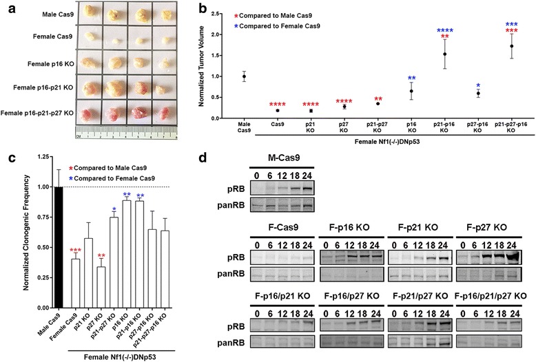
Fig. 6.

Combined loss of p16 and p21 in female GBM astrocytes recapitulates the male GBM phenotype. a Representative flank tumors from male and female GBM Cas9 control astrocyte initiated tumors. b Quantification of mean and SEM tumor volumes of male and female GBM Cas9 control tumors and each of the p16, p21 and p27 single and combinatorial KO female cell lines. Tumors were harvested at 8-weeks post-implantation and measurements are of ex vivo tumors. Statistical significance was determined using either male Cas9 tumors as reference (red asterisks) or female Cas9 tumors as reference (blue asterisks). Asterisks (1–4) refer to p values of < 0.05, < 0.005, < 0.0005, or < 0.00005 as determined by one-way ANOVA and Dunnett’s post-hoc test (n = 15 for p21 KO and p27 KO, and n = 5 for each of p21;p27 DKO, p16 KO, p21-p16 DKO, p27-p16 DKO and p21-p27-p16 TKO). c ELDA assays were utilized to measure clonogenic cell frequency. Asterisks refer to comparisons between male and female Cas9 GBM cells and p-values are as described for panel b. d The effect of p16, p21 and p27 loss on Rb phosphorylation was measured by Western blot analysis of cells stimulated with serum after 48 h of serum starvation. Shown are representative blots from individual experiments
