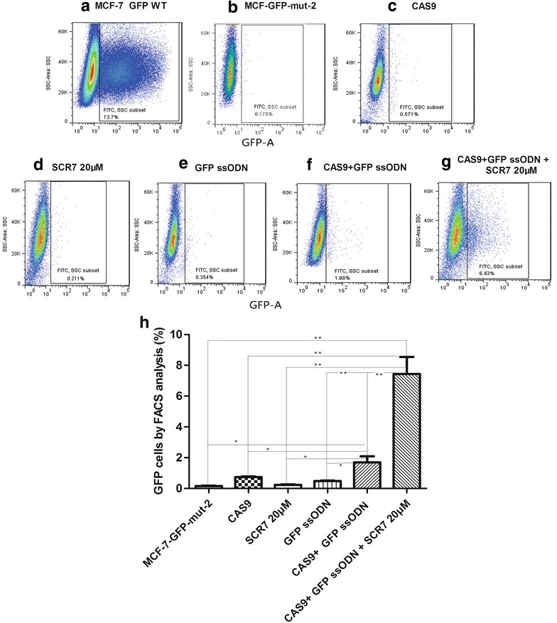
Fig. 5.

The enhancement of mutation-correction efficiency of a GFP-silent mutation by ssODN and SCR7 treatment in MCF-7/GFP-Mut cells. MCF-7/GFP-Mut cells were co-transfected with the pCS2-Cas9-U6-sgRNA (GFP-Mut) vector or/and GFP ssODN. After transfection, cells were treated with vehicle control or SCR7 (20 μM) for 48 h. At the end of experiment, the GFP-positive cells were quantified by FACS. MCF-7 cells transfected with the wild-type GFP vector (pSIN-EF1-GFP-puromycin) were used as GFP-positive control cells. a–g show the representative flow cytometric analyses of MCF-7 cells transfected with the wild-type GFP vector (a), MCF-7/GFP-Mut cells without transfection (b), MCF-7/GFP-Mut cells transfected with pCS2-Cas9-U6-sgRNA (c), or GFP ssODN alone (e), MCF-7/GFP-Mut cells treated with 20 μM SCR7 alone (d), and MCF-7/GFP-Mut cells transfected with both pCS2-Cas9-U6-sgRNA and GFP ssODN without (f), or with 20 μM SCR7 treatment (g). h shows a quantitation of GFP-positive cells in percentage (mean ± SD) from 3 independent experiments. SSC—side scatter. *p < 0.05, **p < 0.01
