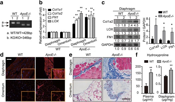
Fig. 2.

ApoE loss leads to altered peritoneal ECM composition. (a) Agarose gel electrophoresis of ApoE gene. (b) The mRNA expression of Collagen1, FN1 and LOX in the excised diaphragm and omentum of WT and ApoE−/− mice. (c) Western blot analysis of Col1a2, LOX and FN1 protein levels in the diaphragm. 1 and 2 represent different samples. (d) Frozen sections were immunoassayed for LOX (red) and nuclei were stained with Hoechst (blue). (e) The representative images of Masson’s Trichrome staining in the diaphragm and omentum from WT and ApoE−/− mice. (f) The plasma and diaphragm were analyzed for hydroxyproline content. Error bars represent the SD of triplicate experiments. Bar represents 50 μm. *P < 0.05; **P < 0.005
