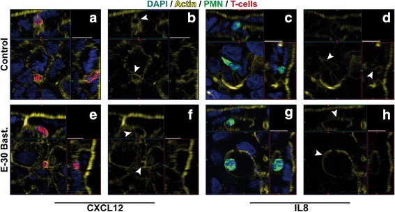
Fig. 6.

Immunofluorescence analysis of leukocyte migration pathway across the HIBCPP. Immune cell migration across HIBCPP monolayers infected with E-30 Bastianni was analyzed using immunofluorescent staining. Infection for 28 h with E-30 Bastianni was carried out before staining the filters for nuclei with DAPI (shown here in blue), actin skeleton with phalloidin (shown here in yellow), PMN (shown here in green), and T-cells (shown here in red). For detailed description of image acquisition and preparation, please refer to Fig. 2. Image pairs show regions of interest and a single channel view of actin expression. White arrowheads indicate the localization of the leukocyte in relation to the actin skeleton. The surrounding of leukocytes by intercellular actin borders which are connected to the apical epithelial cell membrane is in favor of paracellular diapedesis (b, d, f), whereas the localization of the leukocyte in clear distance of the actin skeleton is in favor of transcellular diapedesis (h). Images were taken for each of the following conditions: HIBCPP cells + PMN and T-cells + CXCL12 = a, b; HIBCPP cells + PMN and T-cells + E-30 Bastianni + CXCL12 = e, f; HIBCPP cells + PMN and T-cells + IL8 = c, d; HIBCPP cells + PMN and T-cells + E-30 Bastianni + IL8 = g, h. These are representative images of three independent experiments, each carried out in duplicates and analyzed with a minimum of four z-stacks per filter
