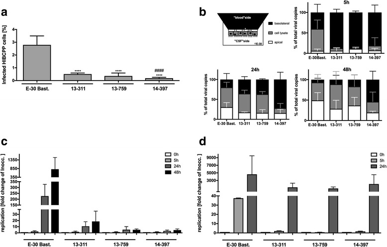
Fig. 9.

E-30 strain-specific variance in replication and viral dissemination in HIBCPP and RD cells. HIBCPP cells or RD cells were infected with E-30 Bastianni, 13-311, 13-759, and 14-397 for up to 48 h (MOI 0.7). a Firstly, filters were stained for DAPI and VP1. Then quantification of infected cells was carried out following the procedure described in the material and methods section. Bars display the percentage of infected HIBCPP cells. Data are shown as mean + SD from three independent experiments carried out in duplicates. p values were calculated for the comparison of E-30 Bastianni-infected HIBCPP cells against infected with either outbreak strain (****p < 0.0001) or for comparison of 13-759 and 14-397 with 13-311 (####p < 0.0001). b The distribution of viral genomes was analyzed by qPCR within the cells and two extra-cellular sides. At each respective time point (5, 24, and 48 h), supernatant from both the filter compartment (basolateral cell side) and the well compartment (apical cell side) was collected. Additionally, filter membranes covered with a HIBCPP cell-layer were lysed to assess cell-associated virus. The upper left shows a schematic view of the different compartments compared in this experiment: the basolateral compartment (black), the cell lysate (gray), and the apical compartment (white). Basolaterally (shown here in black), cell associated (shown here in gray), and apically (shown here in white) located viral particles are represented as percentage of total viral particles for the particular strain. On the upper right the results after 5 h of infection, on the lower left the results after 24 h of infection, and on the right the results after 48 h of infection are shown. Data shown are mean + SD from at least three independent experiments carried out in triplicates. c After infection of HIBCPP cells, collection of cell free (from the filter and well compartments) and cell-associated virus was performed at indicated time points: 0 h (white bars), 5 h (light gray bars), 24 h (dark gray bars), and 48 h (black bars). The measurement of viral genome copies was carried out with qPCR. Data are shown as mean + SD for three independent experiments performed in duplicates (*p < 0.05, ****p < 0.00001). d After infection of RD cells, collection of cell free (from the well) and cell-associated virus particles was carried out at indicated time points: 0 h (white bars), 5 h (light gray bars), and 24 h (dark gray bars). Data are shown as mean + SD taken from four independent experiments carried out in triplicates
