Figure 2.

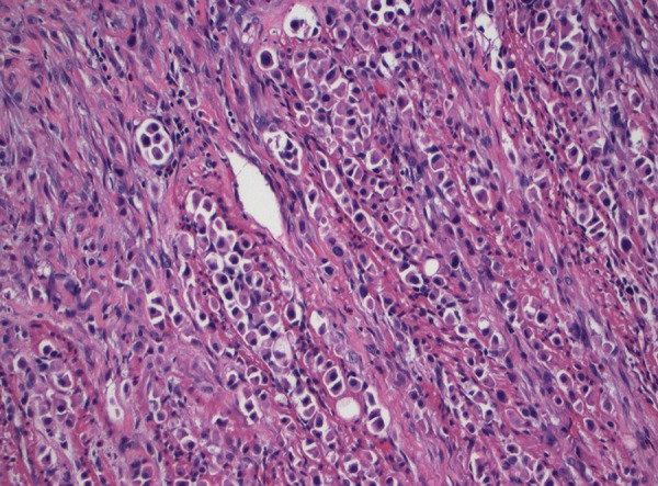
High-magnification pathological biopsy of segment of resected small intestine showing high-grade urothelial carcinoma with plasmacytoid features, characterized by abundant cytoplasm, eccentric nuclei, and discohesive invasive pattern.

High-magnification pathological biopsy of segment of resected small intestine showing high-grade urothelial carcinoma with plasmacytoid features, characterized by abundant cytoplasm, eccentric nuclei, and discohesive invasive pattern.