
Figure 7. 7T images of giant macroadenoma.
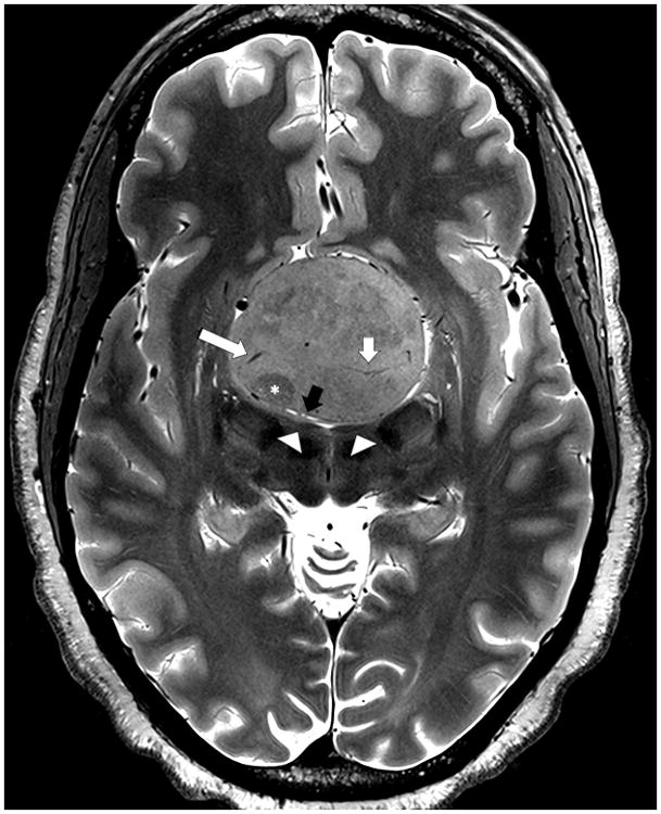
Axial T2-weighted TSE image obtained at 7T in a patient with a giant pituitary adenoma. The image shows tumor heterogeneity including zone of lower signal suggesting a region of firmer tumor (asterisk), venous drainage (long white arrow), tumor arterial supply (short white arrow) and compressed adjacent anatomy including flattened mammillary bodies (arrowheads) and the lamina terminalis (black arrow).