Fig. 2.
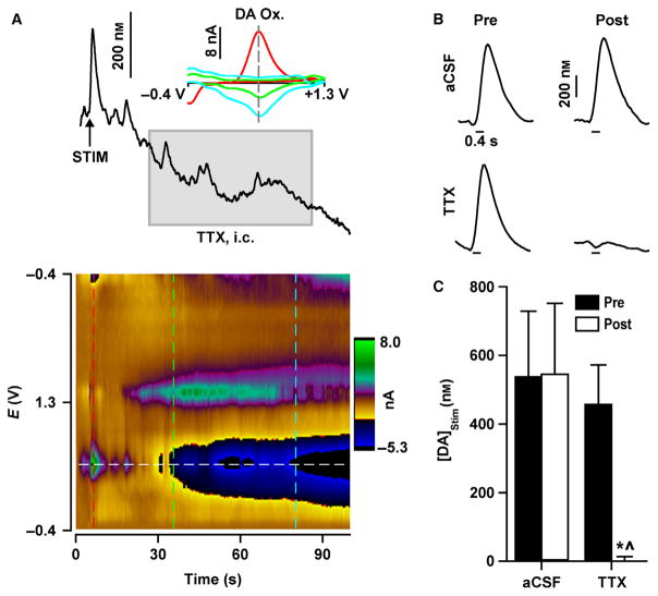
TTX abolishes electrically evoked dopamine signals. (A) Representative trace during an intracranial TTX infusion [grey box; TTX, intracranial (i.c.)] into the VTA. Electrical stimulation (STIM; vertical arrow) applied to the VTA prior to TTX elicited dopamine signals mimicking naturally occurring dopamine transients. TTX infusion caused a decrease in basal dopamine that tracks the change in current determined at the dopamine oxidative potential (DA Ox.), as shown by individual background-subtracted cyclic voltammograms (inset) measured at different time points (vertical dashed lines on colour plot). Similar dopamine decreases were also evident in the colour plot (horizontal dashed white line). (B) Representative electrically evoked dopamine signals (stimulation indicated by horizontal line under traces) before (Pre) and after (Post) aCSF or TTX infusion. (C) Maximal concentration of electrically evoked dopamine ([DA]Stim) was abolished by TTX but unaffected by aCSF. Data are expressed as mean + SEM. *P < 0.01 vs. Pre (within-group comparisons); ^P < 0.01 vs. aCSF (between-group comparisons).

