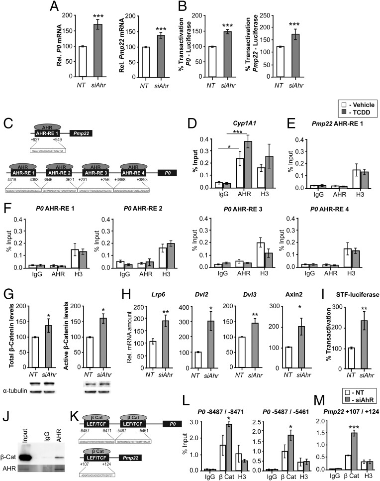
Fig. 5.
AHR does not bind to myelin gene promoters in MSC80 cells but acts via the Wnt/β-catenin pathway. MSC80 cells were transfected with a siRNA directed against Ahr (siAHR) or a NT siRNA (NT). (A) Total mRNA was extracted, and P0 and Pmp22 transcripts were quantified by qRT-PCR. All results were normalized to the 26S mRNA level. (B) MSC80 cells were cotransfected with siAhR or NT and P0-luciferase or PMP22-luciferase constructs. Luciferase activity was assayed. Results represent the means ± SEM of at least five independent experiments. ***P < 0.001 by Student’s t test compared with control (NT). (C) Putative binding sites of AHR located on the levels of P0 and PMP22 were identified by means of MatInspector software. MSC80 cells were incubated with vehicle (Nonane) or TCDD (100 µM, 24 h). ChIP assays with antibodies against AHR or Mock IgG (nonrelevant negative control) or Histone H3 (positive control) were performed. qRT-PCR was performed with primers recognizing AHR putative sites located on (D) Cyp1A1, an AHR target gene; (E) Pmp22; and (F) P0 genes. Results represent the means ± SEM of at least four independent experiments. *P < 0.05 and ***P < 0.001 by Tukey’s post hoc tests after one-way ANOVA compared with control. (G) MSC80 cells were transfected with siRNA directed against Ahr (siAhr) or a nontargeting siRNA (NT) for 48 h. Total proteins were extracted, and WBs were performed using antibodies against total β-catenin or active β-catenin and α-tubulin as a loading control. Figures represent a typical experiment. WB quantifications were done using ImageJ software. (H) Total RNA was extracted, and qRT-PCR experiments were performed using primers recognizing Lrp6, Dvl2, Dvl3, and Axin2. RT-PCR was normalized using 26S RNA. Data represent the mean ± SEM of at least four independent experiments. (I) MSC80 cells were transiently transfected with SuperTOP Flash-luciferase (STF-Luciferase) and with either siAHR or NT. Forty-eight hours later, luciferase activity was analyzed. Results represent the means ± SEM of at least six independent experiments performed in duplicate. *P < 0.05, **P < 0.01, by Student’s t test. (J) Coimmunoprecipitation assays were performed on MSC80 cell extracts using AHR, blotted for β-catenin by WB, and lgG was used as a control. (K) The binding sites for TCF/LEF–β-catenin are localized on the promoter level for P0 and Pmp22 as described in ref. 12. MSC80 cells were transiently transfected with either siAhr or NT for 48 h after; ChIP assays with antibodies against β-catenin or Mock IgG or Histone H3 were peformed. Primers recognizing TCF/LEF–β-catenin binding sites on the levels of P0 (L) and Pmp22 (M) were used. Results represent the means ± SEM of at four independent experiments. *P < 0.05, ***P < 0.001 by Tukey’s post hoc tests after one-way ANOVA compared with control.

