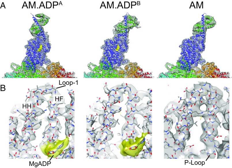
Fig. 1.
Structural states of actin-bound myo1b in the presence and absence of MgADP. (A) Cryo-EM density maps (gray) and final fitted AM.ADPA, AM.ADPB, and AM models. The myo1b protein construct (blue) with a single IQ motif and bound calmodulin (dark green) are bound to actin subunits (cyan, green, light green, orange, and red). Cryo-EM Density for MgADP is highlighted in gold. (B) Cryo-EM density map showing the nucleotide binding site. The MgADP (gold) is bound to the P loop and framed by the HF and HH helices, which are connected by loop 1.

