Fig. 1.
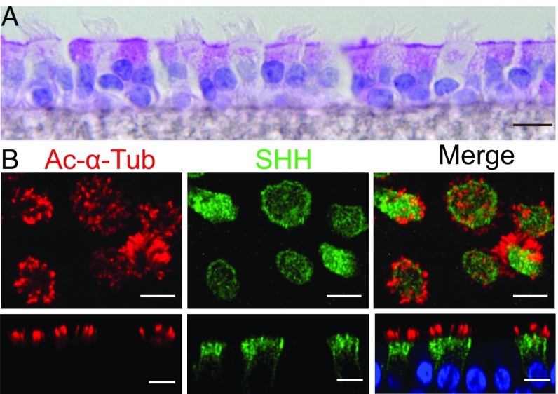
SHH is expressed in ciliated airway epithelial cells. Images in this and other figures are from primary cultures of differentiated human airway epithelia, except in Fig. S4, which is lung tissue. (A) Section of periodic acid Schiff-stained epithelia. (B) Immunostaining of acetylated α-tubulin (red), SHH (green), and DAPI (nuclei, blue). Upper images are stacks of X-Y confocal images, and Lower images are X-Z images. (Scale bars, 10 μm.)

