Fig. 3.
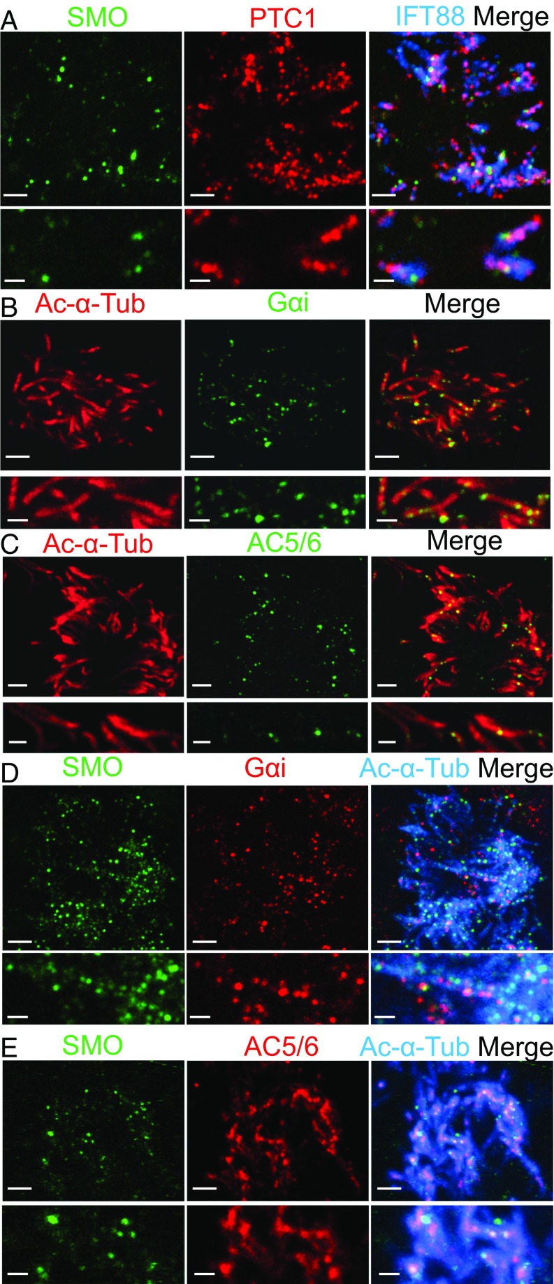
Gαi and AC5/6 are expressed with SMO in motile cilia. (A) Staining of SMO (green), PTC1 (red), and IFT88 (cilia, blue). (B and C) Staining of acetylated α-tubulin is red, and staining of Gαi (B) and AC5/6 (C) is green. (D and E) Staining of SMO is green, staining of Gαi (D) and AC5/6 (E) is red, and staining of acetylated α-tubulin is blue. Lower images are expanded images from Upper. [Scale bars: 2 μm (Upper), 1 μm (Lower).]

