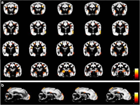
Fig. 4.

Results of VBM analysis 3 (control vs. epileptic Beagle group) using SPM12. Transversal [a] and Sagittal [b] view of parts of the GM-template of control Beagles illustrating regions of significant reduced GMV (coloured voxels) in the olfactory bulb, cortex of frontal, parietal, temporal and occipital lobe, cingulate gyrus, putamen, claustrum, hippocampus and cerebellum in the epileptic Beagle group (VBM: voxel-based morphometry, GM: grey matter, GMV: grey matter volume)
