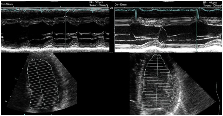
Fig 4. Echocardiography images of M-mode and apical two-chamber views.
Athlete 1 was classified as eccentric LVH with 2TC due to a LVID of 57.1 mm and a PWT of 9.8 mm resulting in a relative wall thickness of 0.34. He was reclassified with 4TC to concentric non-dilated LVH due to a relative short LV length of 8.2 cm and an associated LV EDV/BSA of 60.1 ml/m2 resulting in a Concentricity of 10.4 ml/m2/3. Athlete 2 was also classified as eccentric LVH with 2TC due to a LVID of 53.6 mm and a PWT of 10.6 mm resulting in a relative wall thickness of 0.40. In contrast to Athlete 1, Athlete 2 was reclassified with 4TC to eccentric non-dilated LVH due to a longer LV length of 9.3 cm and an associated higher LV EDV/BSA of 71.5 ml/m2 resulting in a smaller Concentricity of 8.7 ml/m2/3.

