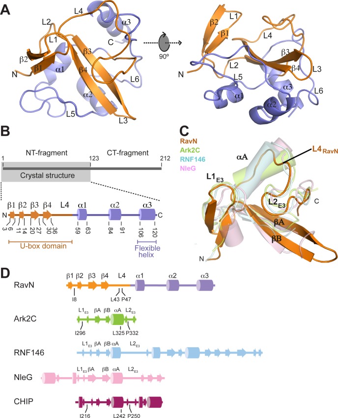
Fig 3. The E3 ligase domain of RavN has a contorted U-box fold.
(A) Schematic representation of the structure of RavN1-123 as ribbon diagram displayed in two orientations (rotated by 90° along the x axis). Secondary elements are indicated as spirals (helices) or arrows (beta strands), with the RING/U-box motif colored in orange and the C-terminal structure colored in slate. (B) Topology diagram of RavN1-123 with the same color scheme as shown in (A). Numbers indicate amino acid residues. (C) Superimposition of the U-box domain of RavN (RavN1-55, orange) with the RING domains of Ark2C (PDB 5D0I, light green) and RNF146 (PDB 4QPL, light blue), and the U-box domain of NleG (PDB 2KKX, light pink). Conserved structural elements shared among RING/U-box domains are labeled as L1E3, L2E3, αA, βA, and βB. The position of αA is occupied by loop L4 in RavN (L4RavN). (D) Topology diagrams of the structural homologs of RavN. The same color scheme is used as in (C) and in Fig 4A. The residues located at the E2 binding interface in RavN, Ark2C, and CHIP (as seen in Fig 4A) are indicated.

