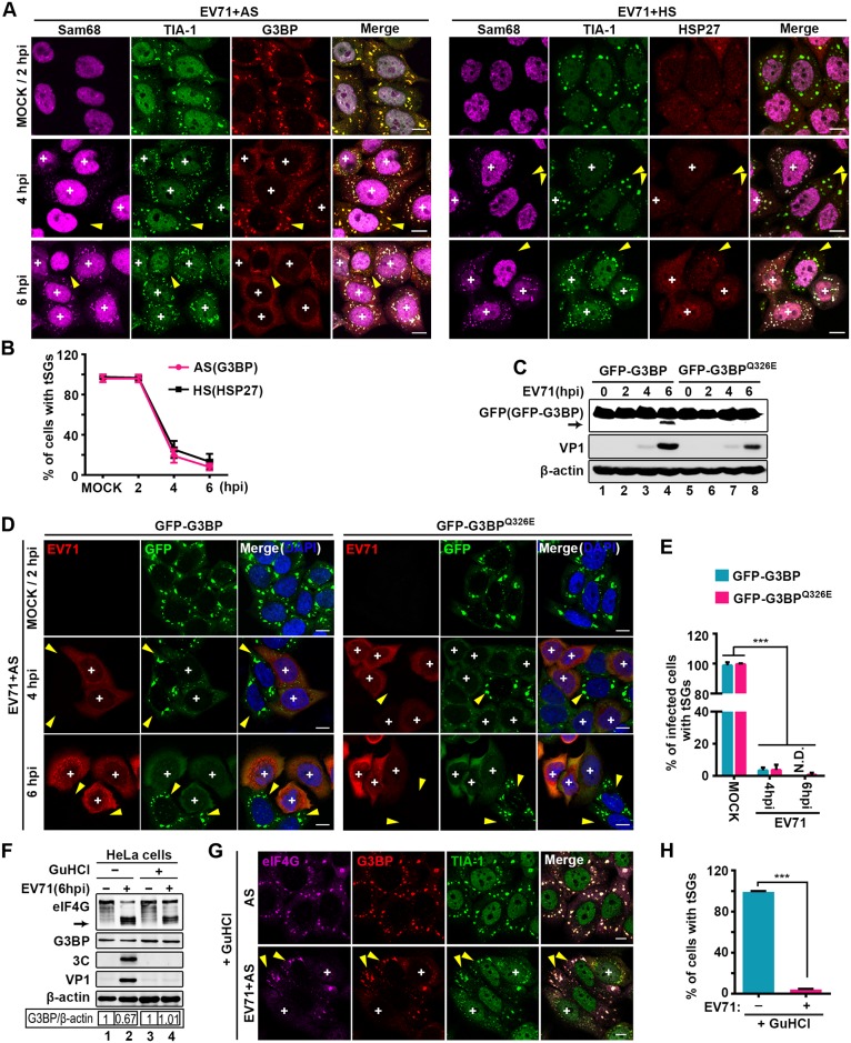
Fig 3. EV71 infection blocks tSG formation independent of 3C.
(A) EV71 effects on AS- and HS-induced tSG formation. HeLa cells were mock-infected or infected with EV71 (MOI = 10) for consecutive times and treated with AS or HS for 1 h prior to fixation. Cells were then stained with Sam68 (magenta), TIA-1 (green), G3BP (red), or HSP27 (red) as indicated. (B) Quantitative analysis of cells with tSGs in A. tSGs were marked by G3BP or HSP27. n = 3, 300 cells/condition were counted, mean±SD. (C) GFP-G3BP- or GFP-G3BPQ326E-HeLa cells were infected with EV71 as in A. Cells were harvested and analyzed via WB. VP1 served as an EV71 marker, and β-actin was the loading control. Full-length GFP-G3BP and N-terminal cleavage products of GFP-G3BP were detected by antibody against GFP, and arrow indicates the N-terminal cleavage products of GFP-G3BP. (D) GFP-G3BP- or GFP-G3BPQ326E-HeLa cells were treated and harvested as in A. Cells were then stained with EV71 (red), GFP-G3BP and GFP-G3BPQ326E (green) were markers of tSGs. (E) Quantitative analysis of EV71-infected cells with tSGs in D. n = 3, 300 cells/condition were counted, mean±SD; ***p<0.001. N.D., not detected. (F) HeLa cells were mock-infected or infected with EV71 in the presence or absence of GuHCl for 6 h as indicated and subjected to WB. Endogenous eIF4G and G3BP were detected by antibodies against eIF4G and G3BP. 3C and VP1 expression served as markers of EV71; β-actin was the sample loading control. (G) HeLa cells were mock-infected or infected with EV71 in the presence of GuHCl for 6 h and treated with AS for 1 h before harvest. Cells were then stained with eIF4G (magenta), G3BP (red), and TIA-1 (green). (H) Quantitative analysis of EV71-infected cells with tSGs (marked by G3BP) in G. n = 3, 240 cells/condition were counted, mean±SD; ***p<0.001. “+” indicates the EV71-infected cells, and yellow arrows indicate the uninfected cells. Scale bars, 10 μm. See also S4 Fig.

