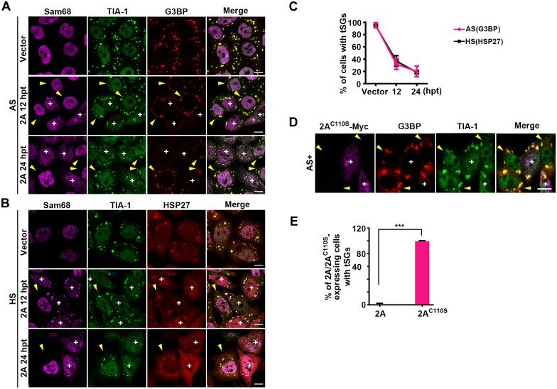
Fig 4. 2A protease can block tSG formation.
(A and B) HeLa cells were transfected with 2A for 12 h and 24 h or transfected with empty vector for 24 h as a control, followed by 1 h treatment with AS or HS, and stained with Sam68 (magenta), TIA-1 (green), G3BP (red), or HSP27 (red) as indicated. (C) Quantitative analysis of cells with tSGs in A and B as marked by G3BP or HSP27. n = 3, 240 cells/condition were counted, mean±SD. (D-E) Effects of protease activity of 2A on tSG blockage. HeLa cells were transfected with Myc-tagged 2AC110S for 24 h and treated with AS for 1 h. tSG formation was viewed by IF assay (D). Quantitation analysis of 2A-expressing cells (in A, 24 hpt) or 2AC110S-expressing cells (in D) with SGs. n = 3, 240 cells/condition were counted, mean±SD; ***p<0.001 (E). “+” indicates the 2A/2AC110S-expressing cells, and yellow arrows indicate the cells without 2A/2AC110S expression. Scale bars, 10 μm. See also S5 Fig.

