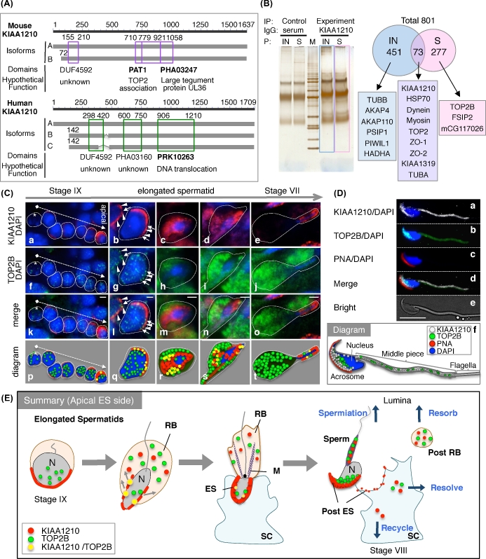
Figure 4.
Characterization and localization of KIAA1210 with TOP2B in the testis. (A) The isoforms and domains of KIAA1210, two isoforms (line A, B) in mouse and three isoforms (line A, B, and C) in human were identified. Purple or green box, domain; number, the number of amino acids from N-terminus. (B) Immunoprecipitation experiments using proteomics analysis (IP-proteomics). Immunoprecipitation using anti-KIAA1210 antibody (KIAA1210) and serum as control (serum) to insoluble (IN) or soluble (S) testis lysate was performed, run to SDS-PAGE gel, and suffered silver stain (left). M, protein marker. The proteomics date of blue and pink opened box in left of Figure 4B (right). Arabic numbers, number of identified proteins. (C) Localizations of KIAA1210 (red) and TOP2B (green) with DAPI (blue) in the testis. Immunofluorescence images of KIAA1210 (a–e), TOP2B (f–j), merged (k–o), and diagram (p–t) in stage IX seminiferous tubule (a, f, k, p), surrounding area of elongated spermatids (b–d, g–i, l–n, and q–s), and stage VII seminiferous tubule (e, j, o, t) are shown. Asterisk, nonexpression cell of KIAA1210; dotted arrow, from basal to apical; arrow, strong co-localization of KIAA1210 and TOP2B; arrowhead, TOP2B on weak signal of KIAA1210. Bar, 2 μm. (D) Subcellular localization of KIAA1210 and TOP2B in spermatozoa. Immunofluorescence images of KIAA1210 (a, white), TOP2B (b, green), PNA (c, red), merged (d), and bright field (e) are shown. A diagram of the merged image is shown in panel f. Bar, 10 μm. (E) The summary of dynamic change of KIAA1210 and TOP2B during spermiogenesis. N, nucleus; ES, apical ectoplasmic specialization; RB, residual body; SC, Sertoli cell; M, manchette.

