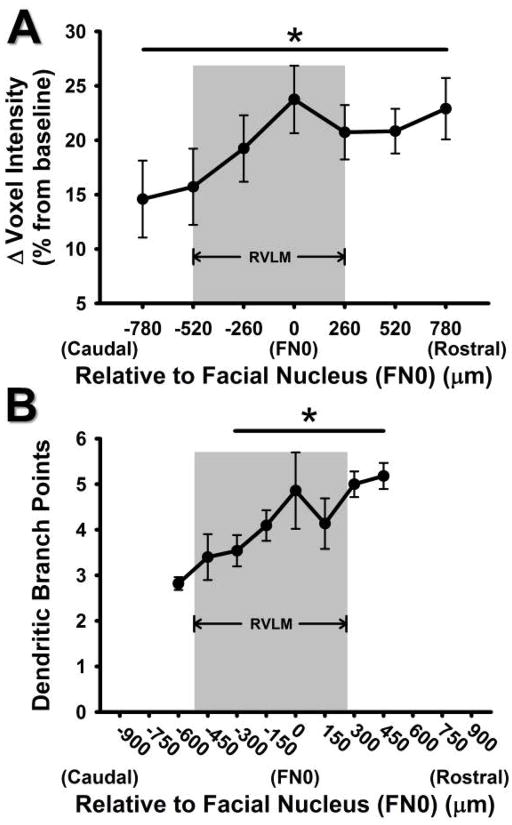
Figure 4. Comparison of caudal to rostral change in voxel intensity in the current study versus caudal to rostral change in dendritic branching from our previous study.
A) Percent change in ROI mean voxel intensities of Mn2+ 24 hours after injection of 66 mg/kg MnCl2, using T1 weighted MRI scanning. The x-axis represents caudal to rostral axis of the ventrolateral medulla relative to the caudal pole of the facial nucleus (designated as FN0 and 0 μm). A pattern of increasing Mn2+ uptake was seen from the caudal to rostral levels of the RVLM (*, p<0.05, main effect). B) Pattern of increased dendritic branching from caudal to rostral areas of the ventrolateral medulla observed in bulbospinal catecholaminergic RVLM neurons from our previous study (*, p<0.05, compared to 600 μm; modified from Mischel et al., 2014). Grey boxes represent the functionally and anatomically defined region of the RVLM.

