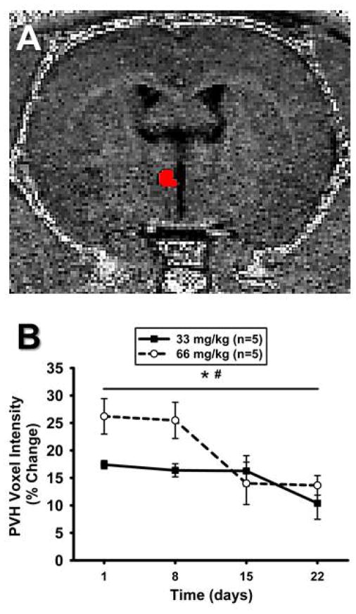
Figure 5. Mn2+ enhancement in the PVH over time using T1-weighted imaging.
A) Unprocessed, non-colorized T1-weighted MR image from one animal demonstrating detailed anatomy. ROI depicted in red and was used to determine voxel intensities in the PVH. B). Time course representing percent change in ROI mean voxel intensity in the PVH following systemic injections of MnCl2 at Day 0 (not shown) and calculated as a percent change from non-Mn2+-injected baseline scans. ROI mean voxel intensity was highest at 24hrs and significantly decreased over time in both groups (*, p<0.05 main effect of time). ROI mean voxel intensity was greater in animals injected with 66 mg/kg, compared to 33 mg/kg MnCl2 (#, p<0.05 main effect of dose).

