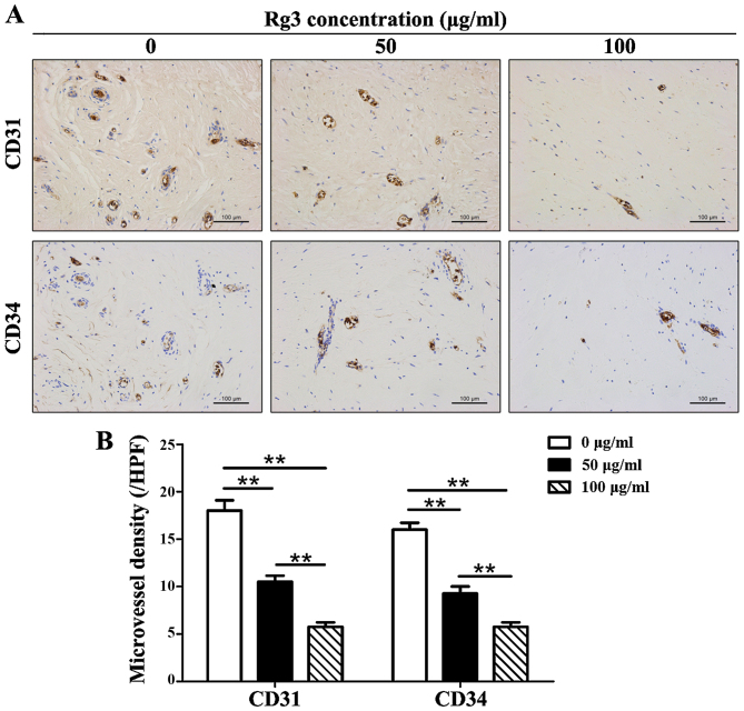
Figure 6.
Immunohistochemical analysis of CD31 and CD34 in keloid explant cultures. (A) Number of CD31 and CD34 positively stained microvessels was decreased in Rg3-treated groups (scale bar, 100 μm). (B) Quantitative analysis of immunohistochemistry indicated that microvessel density was reduced by ~1/2 in the 50 μg/ml-treated group and by ~2/3 in the 100 μg/ml-treated group compared with in the control group. **P<0.01. CD, cluster of differentiation; Rg3, ginsenoside Rg3.

