Figure 2.
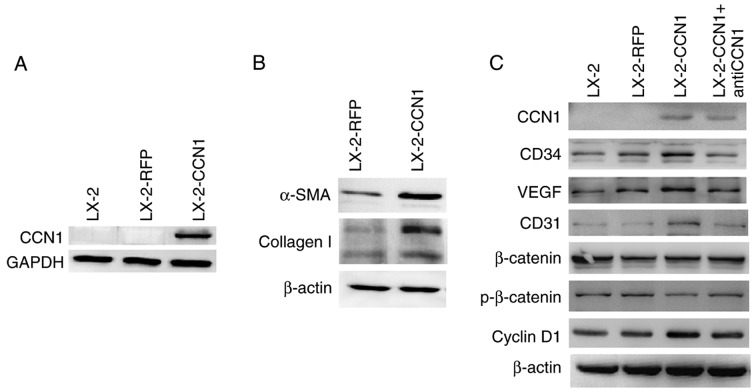
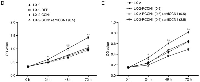
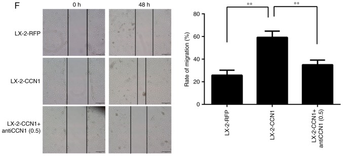
CCN1 activates HSCs and affects cell function. LX-2 cells were infected with adenovirus AdCCN1 or AdRFP, respectively. (A) Following infection for 24 h, western blot analysis was performed to detect the expression levels of CCN1 in LX-2 cells, LX-2-RFP cells and LX-2-CCN1 cells. (B) Following infection for 72 h, α-SMA and collagen I, which are markers of HSC activation and fibrosis, were detected in the LX-2-CCN1 cells. (C) Expression levels of p-β-catenin, β-catenin, cyclin D1, VEGF, CD31 and CD34 were analyzed. CCN1 antibody (0.5 μg/ml) was used to neutralize CCN1 in control groups. (D) Viability of LX-2-CCN1 cells was analyzed using MTT assays. (E) Following treatment with RCCN1 (0.6 μg/ml), LX-2 cells were analyzed using MTT assays. CCN1 antibody (0.5 mg/ml) was used in assays. The final concentrations were 0.5 and 2.5 μg/ml. (F) Monolayer scratch assay was used to analyze the migration of LX-2-CCN1 cells. *P<0.05 and **P<0.01. HSCs, hepatic stellate cells; CCN1, cysteine-rich 61; VEGF, vascular endothelial growth factor; SMA, smooth muscle actin; p-, phosphorylated; RCCN1, recombined CCN1; OD, optical density.


