Figure 5.
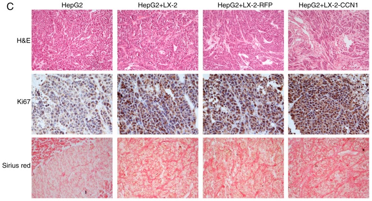
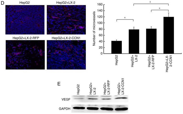
CCN1 enhances the function of hepatic stellate cells in promoting the proliferation of hepatocellular carcinoma cells in vivo. (A) Nude mice and tumor tissues of animals following injection at 26 days. (B) Tumor sizes were measured at a 4-day interval from day 6 post-injection. The graph shows the tumor volumes for each treatment group. *P<0.05 and **P<0.01 vs. the CM-LX-2-RFP group. (C) H&E staining of tumor tissues showed hyperplasia of fibrous connective tissues, inflammatory cell infiltration and multinuclear tumor cells in subcutaneous tumor samples (magnification, ×200). Immunohistochemical detection of Ki67 in subcutaneous tumor samples (magnification, ×200). Collagenous fibers in subcutaneous tumor were observed using Sirius red staining (magnification, ×200). (D) Microvessels in subcutaneous tumor tissues were observed using immunofluorescence staining for CD31 (magnification, ×200). Data were quantified as the number of microvessels (CD31-positive endothelial cells). *P<0.05. (E) Protein expression of VEGF in subcutaneous tumor tissues was detected using western blot analysis. CCN1, cysteine-rich 61; H&E, hematoxylin and eosin; VEGF, vascular endothelial growth factor.


