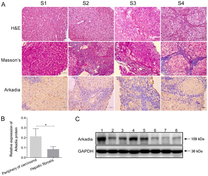
Figure 5.
Arkadia expression in fibrotic liver samples from human patients. Liver samples were obtained from patients with hepatic fibrosis at various stages, and underwent H&E and Masson staining. The protein expression levels of Arkadia were detected in fibrotic and nonfibrotic liver samples by immunohistochemistry and western blot analysis. Results are presented as representative images or are expressed as the means ± standard deviation of individual groups. (A) Histological and immunohistochemical examination of liver sections (original magnification, ×200). S1, S2, S3 and S4, hepatic fibrosis at the indicated stage. (B) Relative protein expression levels of Arkadia. (C) Western blot analysis of the protein expression levels of Arkadia. Lanes 1–5, periphery of carcinoma; lanes 6–8, hepatic fibrosis. *P<0.05. H&E, hematoxylin and eosin.

