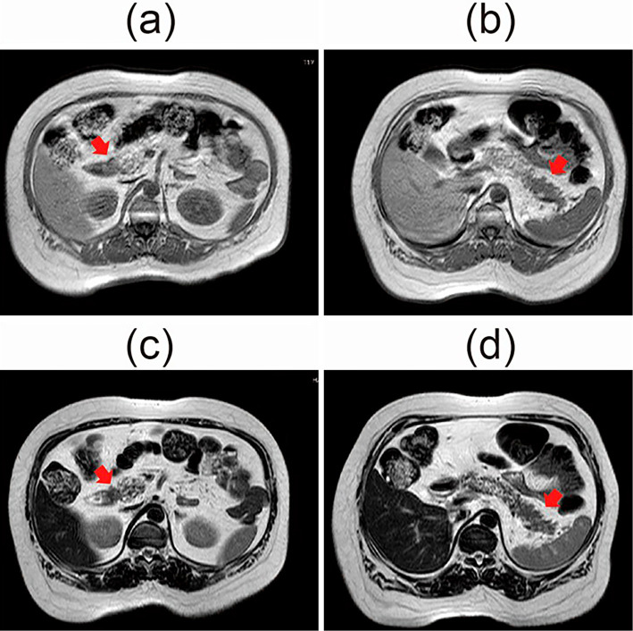
Figure 2.
Magnetic resonance imaging (MRI) revealed the presence of pancreatic lesions. These pancreatic lesions showed slightly low intensity on T1-weighted images (a: head, b: tail) and slightly high intensity on T2-weighted images (c: head, d: tail) compared to the pancreatic parenchyma and the liver.

