Figure 2.
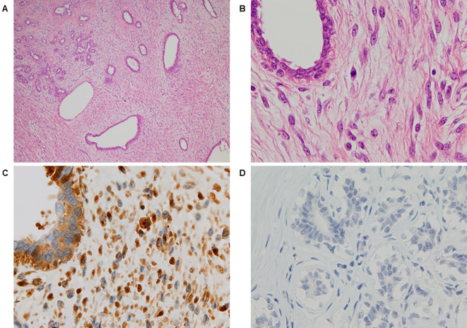
The microscopic findings [Hematoxylin and Eosin (H&E) staining and immunohistochemical staining of the resected tumor]. H&E staining (A) Low-power (×40) and (B) high-power (×400) views showed moderate stromal hypercellularity with mild nuclear atypia and mild pleomorphism of the spindle cells. Focal mildly atypical epithelial hyperplasia was also noted. Stromal overgrowth was absent. Immunohistochemical staining of (C) the phyllodes tumor and (D) normal breast tissue using rabbit polyclonal anti-insulin-like growth factor II (IGF-II) antibodies. The tumor cells, but not the normal breast tissue, were diffusely immunopositive for IGF-II.

