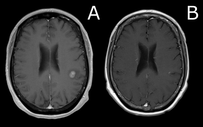
Figure 2. Comparison of brain MRI at presentation versus three months after SRS procedure.
A) MRI brain imaging revealed two contrast-enhancing lesions in the left frontal lobe (pictured in A); T1 axial image, post-gadolinium contrast) and left cerebellar hemisphere, measuring 1.25 cc and 0.07 cc, respectively. The patient underwent stereotactic radiosurgery (SRS), with 22 Gy delivered to the 50% isodose line to each lesion. B) Three months after SRS, repeat MRI demonstrated resolution of the frontal lobe lesion.
MRI: magnetic resonance imaging

