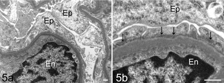
Fig. 5.
Electron micrograph of a glomerulus of the male Sprague Dawley rat with minimal change disease. This rat showed extensive foot process effacement of the podocytes (a; Bar = 50 nm). The slit pore, which is situated between the foot processes, is narrowed, and rearrangement of the actin cytoskeleton (arrows) is observed in fused foot processes in the podocyte (b; Bar = 50 nm). Ep, glomerular epithelial cell (podocyte); En, endothelial cell.

