Fig. 3.
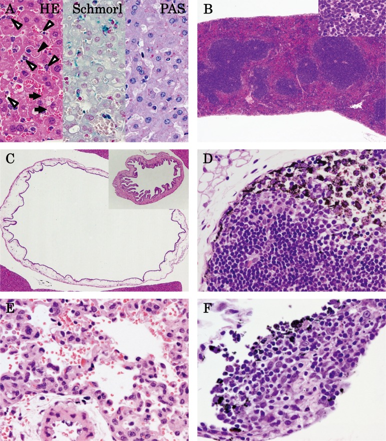
Histopathology of 10 nm AgNPs at 6 h (A–D) post administration, 60 nm AgNPs at 1 h (E) post administration, and 100 nm AgNPs at 6 h (F) post administration. (A) Congestion in the liver, hepatocyte vacuolation and dark brown pigment deposition in Kupffer cells (open arrowhead), hepatocyte single cell necrosis (closed arrowhead), and inclusion bodies in cytoplasm (arrow) were observed in HE staining; a slightly blue reaction of pigments was observed with Schmorl’s stain, but the results were negative with PAS stain (×400). (B) Congestion in the red pulp and apoptosis in the white pulp of the spleen (insert; ×40). (C) Submucosal edema and dilatation in the gall bladder (insert, control; ×40). (D) Dark brown pigment deposition in the medullary sinus and subcapsular sinus of the thoracic lymph node (×400). (E) Hemorrhage in the lung (×400). (F) Dark brown pigment deposition in inflammatory cell foci of the mesenterium (×400).

