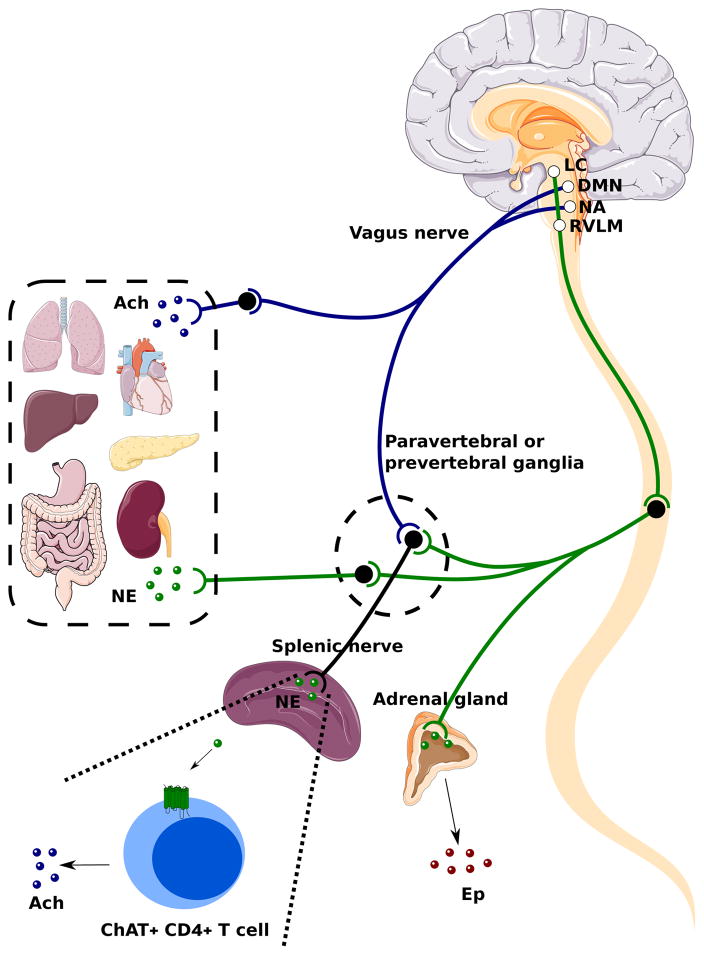
Figure 2. Vagal Efferent Anatomy and the Cholinergic Anti-inflammatory Reflex.
Visceral organs in the thoracic and abdominal cavity including the lungs, heart, liver, stomach, gut, pancreas and kidneys are innervated by parasympathetic and sympathetic nerves. The efferent portion of the vagus nerve, which is the preganglionic parasympathetic fiber, originates from the dorsal motor nucleus (DMN) and nucleus ambiguous (NA) in the medulla oblongata and project to postganglionic fibers in proximity with or within visceral organs. Both the preganglionic and postganglionic vagal fibers are cholinergic. Postganglionic vagal fibers release acetylcholine (Ach) into viscerl organs to regulate smooth muscle activity, gut motility, and gland secretion. In the spinal cord, sympathetic neurons received descending projects from the locus coeruleus (LC) and the rostroventrolateral medulla (RMLM) in the brainstem. The sympathetic preganglionic fibers interact with postganglionic fibers within the paravertebral and prevertebral ganglia. Postganglionic fibers from paravertebral and prevertebral ganglia innervate visceral organs in the thorax (lungs, heart) and the abdominal cavity (liver, stomach, gut, kidneys, pancreas) respectively, and release norepinephrine (NE). Sympathetic preganglionic fibers also innervate the adrenal medulla directly and stimulate the secretion of epinephrine (EP) from chromaffin cells. Both vagal and sympathetic preganglionic fibers give projections to the splenic nerve within the celiac and superior mesenteric ganglion. In a cholinergic anti-inflammatory reflex, NE released by the splenic nerve acts on choline acetyltransferase (ChAT)+ CD4+ T cells through β2-adrenergic receptors. These T cells release Ach to regulate macrophage production of TNF-α and other cytokines.

