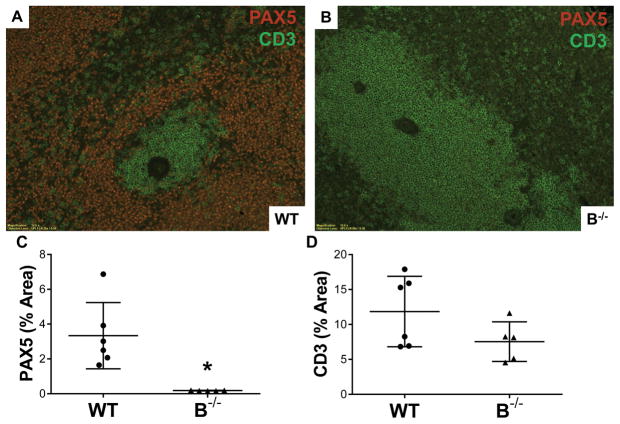
Figure 6. B cell deficient animals lacked splenic germinal centers at 7 days posttransplant.
A, Immunofluorescent staining for both B cells (red, PAX5+) and T cells (green, CD3+) in splenic tissue demonstrated typical germinal center formation in WT. B, B−/− animals lacked B cells and germinal centers. C, Quantitative immunofluorescence demonstrated absence of B cells (PAX5+) in spleens from B−/− rats. * P = 0.005. D, Numbers of splenic T cells (CD3+) were similar between WT and B−/−.

