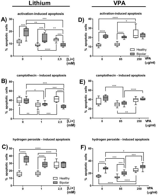
Figure 5.
In vitro influence of lithium carbonate and valproic acid on the percentages of apoptotic T lymphocytes. Figures show percentages of apoptotic cells as annexin-V-positive cells. Apoptosis of lymphocytes was induced either by activation (through stimulation with concanavalin A) (A), camptothecin (B) or hydrogen peroxide (C) alone or in the presence of 1 or 2.5 mM lithium (Li+) in healthy people and BD patients. Meanwhile Figures (D,E and F) show the same parameters in the presence of 85 or 250 µg/ml valproic acid (VPA). Center lines of figures present medians, boxes present 25th–75th percentiles and whiskers show the minimal and maximal values observed, Mann-Whitney test, Friedman ANOVA and Post Hoc, *p < 0.05, **p < 0.01, ***p < 0.001, ****p < 0.0001.

