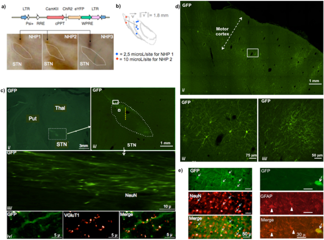
Figure 2.
Histological characterization of the lentiviral vector enabling retrograde transport of ChR2. (a) EIAV-Rabies-CaMK2-ChR2-eYFP and EIAV-VSVg-Rabies-CaMK2-ChR2-eYFP lentiviral vectors with retrograde transfer properties were injected unilaterally in the STN of NHP 1 & 2, and NHP 3 respectively. Cannula traces were visualized on histological slices and confirmed to end above the dorsolateral aspect of STN of NHP 1, 2 & 3. (b) Injection sites for NHP 1 & 2 are 3D-reconstituted in a common STN after affine and elastic transformations between the two STN for NHP 1 & 2. (c) i/ high magnification on a coronal slice showing right STN, thalamus and putamen. ii/ white rectangle around STN on i/ is zoomed. White dots dlimit the STN. Yellow dashes indicate the recording electrode trajectory in the prolongation of the cannula. iii/ the biggest white rectangle in ii/ is zoomed: axons expressing ChR2-eYFP arriving to STN. iv/ the smallest white rectangle in ii/ is zoomed: VgluT1 positive cortico-subthalamic axon terminals expressing ChR2-eYFP. (d) i/ zoom on a coronal motor cortex slice. ii/ white rectangle in i/ is zoomed: neurons of cortical layer V express ChR2-eYFP on their somas, dendrites and initial portion of their axons. iii/ dendrites expressing ChR2-eYFP. (e) Double immune-staining GFP/NeuN and GFP/GFAP in the layer V of motor cortex. White arrows indicate GFP-positive cells. White triangles indicate GFAP-positive cells NHP: Non-human primate; STN: subthalamic nucleus; Thal: thalamus; Put: putamen; VgluT1: vesicular glutamate transporter 1.

