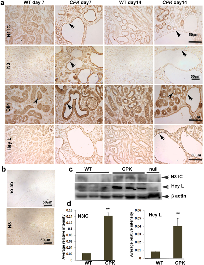
Figure 1.
Expression pattern of Notch pathway members in kidneys of ARPKD mouse model: (a) Immunohistochemistry (IHC) for N1ICD (Notch1 intracellular domain), N3 (Notch3), Dll4 (Delta like 4), and Hey L was performed on paraffin sections of P7 and P14 WT and cpk kidneys. Arrows point to expression in cyst-lining epithelial cells. Arrowheads in third row point to non-cystic tubular cells with Dll4 expression. Images shown are representative of three independent experiments performed in duplicate. (b) Upper panel represents a no primary antibody control. Lower panel shows IHC for N3 on N3-null mouse kidney section to verify antibody specificity. (c) Western Blot for N3IC and Hey L on lysates of P15 WT and cpk kidneys (n = 3), and of N3-null mouse kidneys to verify antibody specificity. (d) Quantitation of WBs for N3IC and Hey L. **P < 0.01.

