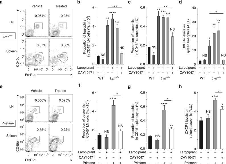
Fig. 6.
PTGDR blockade dampens basophil accumulation in SLO in a lupus environment. a, e Representative contour plots of basophils among living CD45+ cells in LN and spleen from aged Lyn–/– mice (a) and pristane-injected WT mice (e) treated or not (vehicle) with both PTGDR-1 and PTGDR-2 antagonists for 10 days. Proportions are shown on the plots. b−d Comparisons between aged wild-type (WT) treated (n = 4) or not (n = 5) with both PTGDR antagonists and aged Lyn–/– mice treated or not (n = 12) with Laropiprant (n = 5), CAY10471 (n = 4), or both antagonists (n = 9). b, c Proportion of basophils among living CD45+ cells in LN (b) and spleen (c). d CXCR4 expression on spleen basophils in mice as in (c). f–h Comparisons between aged WT mice 24 weeks after injection of either PBS or pristane and treated or not with both PTGDR antagonists (from left to right: n = 12/4/9/4). f, g Proportion of basophils among living CD45+ cells in LN (f) and spleen (g). h CXCR4 expression on spleen basophils in mice as in (f). b–d, f–h Data were determined by flow cytometry as described in Supplementary Fig. 4a–c and represent two pooled independent experiments for each model. Data are presented as mean ± s.e.m. Statistical analyses were by unpaired Student t tests. NS: not significant, *P < 0.05, **P < 0.01, ***P < 0.001, ****P < 0.0001. Comparison to control group is shown above each bar and to the corresponding bars when indicated. A.U. arbitrary units

